Therapies in The Micro-invasive Osteopathy Hospital of TCM

Treatments in The Micro-invasive Osteopathy Hospital of TCM are mainly divided into four methods: “Micro-stitch surgery,” “Chinese Medicine” and “Acupuncture” and “Cupping.”

Micro-surgical Acupuncture: Acupuncture is a micro-surgical procedure originated by Professor Huang Shu, combined with modern Western medicine and traditional Chinese medicine acupuncture anatomy, only with a pin of the TCM operation implemented.

Chinese Herbal Medicine includes: medicine, relieving drugs, Qingre drugs, temperature in medicine, Qi drugs, drug Xiaoshi close, Shibuya medicine, medicine Huashi aromatic medicine, medicine diuresis, Ping asthma drugs, sedative drugs, medicines wind Pinggan intake, remove silt Huoxue drugs, hemostatic, diarrhea medicine, de-worming medicine and aromatic Kaiqiao medicine.

Acupuncture focuses on the fourteen (14) Meridian points in the human body, which are the commonly used acupuncture points, according to the different conditions and different points and select a different way into the needle and the depth and perspective. The fourteen (14) channels are as follows: Governor Vessel, Lung Taiyin hand, Hand Shaoyin Heart Sutra, by the Hand-Jueyin hand Yangming Large Intestine, Small Intestine hand the sun, of Hand-Shaoyang Sanjiao, Stomach Meridian of Foot-Yangming , Full sun, as the bladder, gall bladder by foot Shaoyang enough Taiyin Spleen, Kidney Shaoyin adequate enough Jueyin liver.

Cupping Therapy is a cup-like device using heat as a means of absorbing negative pressure in the adsorption of the acupuncture points on the skin, causing local congestion, congestion as a method of treating disease.

Miracles – Desperate Patients see hopes from TCM in China

What is miracle? Following are medical miracles!

————————————————————-

1. A 48 years old USA Gentleman living in San Fansico:

An accident resulted in the numbness below his neck for 7 years. It was diagnosed as Cervico-vertebral disease. He tried treaments from hospitals all over the World, but the treating results are frustrating. After he got Acupuncture Microsurgery, he completely got recovery. When he was discharged from our hospital, he was very exciting and wrote a Thanks letter.

2. She sought treatments from all famous hospitals in USA she knows, but all told her to “go home”.

It is TCM Acupunture micro-surgery that brought her to normal life. This is one of the TCM miracles which happens every day.

Ms MOSER was desperate because she had failed Surgery of Lumbar Inter-Vertebral Disc Protrusion in USA. However she see hopes from TCM Acupuncture microsurgery.

After the TCM Acupuncture microsurgery, she gave her doctor a thumbs-up sign , said :

“It’s amazing !It’s amazing!It’s amazing!”

这是美国腰椎间盘突出症患者MOSER 女士接受中医针法微创手术后的感慨!75岁的MOSER 女士15年前不幸发生车祸,腰部严重受伤,在美国某著名医院做了腰部的开放手术。开刀手术的损伤程度实在让人恐怖!长长的几处术后瘢痕清晰可见,其中之一是在髋部的瘢痕。手术需要开一个约5厘米的口子,取出正常的骨头,再开个约15厘米的大口子,从腰椎上植入这些骨头加强局部的强度;在椎间盘内植入两个金属的人工椎间盘;骨头上镶嵌了钢钉,里面钉了几枚钢钉,放上了钢板固定。术后,麻药劲儿过MOSER就感到伤口带来的疼痛,那种钻心的痛让她没世不忘。在这样重装手术及漫长休养后,本以为应该解除病痛的MOSER 女士却没有如常所愿。腰痛地还是不能伸展,反而慢慢的出现了驼背,右腿只要迈步,就如刀割肉样疼,根本不敢行走,就连翻身也痛苦难当,经常夜不成寐,一到阴雨寒冷季节就感觉双腿冰冷麻木,上下楼也需要家人搀扶。当MOSER再次回到医院需求医生帮助时,医生的回答是手术只能解决到这种程度了,不过现在还有一种止痛新技术“植入式神经刺激传感器”你不妨试试,也许可以治疗你的疼痛。MOSER查询了一些资料发现植入式神经刺激传感器简称INS2,整套装置有火柴盒那么大,植入到患者的脊椎旁,对神经信号进行监测。一旦发现疼痛信号,就立刻发出电子脉冲,阻止疼痛信号传递到大脑,从而起到缓解疼痛的作用。它是由一个内置的电脑处理器进行操作,动力源则是一块SIM卡大小的电池,进行无线充电。针对不同程度的疼痛,运用自己的“智能”进行“微调”。MOSER认为把一个火柴盒大小的异物放在自己的身体里,使用的装置还需要电池来支持,如果没有及时充电,这个装置也就没有了意义;原理还是是用电子脉冲刺激来阻止疼痛感传递到大脑,要时时注意脉冲的大小,以访导致过大过小影响疗效;总感觉会像一个定时炸弹在身体里随时爆炸一样,手术还需要开刀,之前的手术经历已经让她痛苦不堪,她不像在次承受这种痛苦,所以拒绝了这个手术。本来能歌善舞的MOSER女士竟然整日躲在屋里,郁郁寡欢。MOSER在美国遍求名院名医,可医生们都束手无策,结论都是:(Please go home)回家吧。MOSER女士彻底绝望了。
15年后,一个中国台湾的朋友探望MOSER时提到了中国的微创骨伤中医医院,这家医院,创立了一种不用开刀的中医针法手术,还治愈了自己多年的顽固、严重的颈椎病。当时在医院中也看到过开刀手术失败的患者用这种不用开刀的针法手术治愈,建议她也来试一试。MOSER说:“我痛苦了15年了,也跑遍各大医院,每次都是充满希望的去,失望的回来,一次次的失望让我心灰意冷,感觉我的的世界从此失去了色彩。我常常在想哪怕能减轻我10%的痛苦,我也就感恩戴德了,我也清楚我的病太重了,也太难治了。听你这么说也许这个医院就是我最后的希望了。”当时我真希望能马上来北京治疗,但是等待看黄枢教授门诊的病人太多,我也只能排队等待。
等待的日子如同度日如年,终于MOSER等到了医院的通知,来到了中国。专家们听完MOSER 女士的阐述后,为她做了仔细的会诊检查。因为MOSER 是开刀失败的患者,专家们要根据MOSER 女士的自身情况与影像学检查结果,为她量身定做治疗方案。教授们看到她对手术还是愁眉不展,就告诉她:“我对你的病有信心,因为我们已经治疗了不少这样的患者,像你这样的情况,在德国我也经常为这类患者手术,至少能为你减轻痛苦,也请你对自己和我们有信心!”。MOSER的眼中闪出希望的光亮!

MOSER在美国做手术时,美国的医生给她开刀,首先取出正常的髂骨,但是开刀给她造成的创伤的刀口,长约5厘米

MOSER在美国做腰椎开放手术后的15厘米长的伤疤

MOSER 女士的影像学检查

MOSER 女士的影像学检查订的金属钉、装了2个金属人工椎间盘。

MOSER 女士的影像学检查

院长亲自为MOSER 女士做检查

3月14日上午,由黄院长亲自为MOSER 女士做了中医针法(黄帝针)手术,手术历时32分钟顺利结束。手术中黄院长不断地问MOSER女士的感觉,一步步小心翼翼的把粘连的神经分离,把压迫的组织转移开,动作虽然轻柔但准确有功力。术后MOSER 女士忘乎所以地说道:“我之前在美国做西医开刀手术是全麻,当时躺在那里没有知觉,没想到麻醉过后钻心的疼就一下出来了,而这次手术居然是局部麻醉,我真没想到,原来这么大型的手术还可以局部麻醉。这次手术过程中也没有太大的痛苦,就是打麻药进针时和分离粘连时有一点点痛,黄教授说这是因为我的粘连太重了,要是一般的腰椎间盘突出症的患者,根本是不会感到痛的。手术中,医生也随时与我沟通,问我进针后有没有什么反应和感觉,让这个手术更加安全,因为医生可以时刻的了解我的感受。我先前经历过开刀手术,被告知做了2个小时,这次手术我没觉得有多久,医生就告诉我手术成功结束了,我当时很惊讶,不可思议呀,手术时间也太短了,真是神奇;最让我吃惊的就是仅仅用了粗一点的针就解决了我的问题。这也太神奇了。”

黄院长正在为MOSER 女士进行中医针法(黄帝针)手术

黄院长正在为MOSER 女士进行中医针法(黄帝针)手术

MOSER经过黄院长的中医针法(黄帝针)手术治疗后的针眼

次日,MOSER 女士就感到疼痛减轻大半,腰痛已经消失了,腿上的疼痛已经不到1/3了,并且整日感觉右腿在发热,力量一天比一天强,晚上睡觉翻身轻松多了, 连腰都挺直了。
为了能更好的了解MOSER的术后反应情况,医院专门派遣英语流利的张医生每日与其进行医疗沟通。医院每日要完成10台以上的大型手术,近100台小型手术,护士们忙碌得很,但王素珍护士长强调人性护理,护理不能单独针对医疗,还要针对患者的具体生活饮食,MOSER就是需要特殊对待的一位,由于语言障碍及身体的特殊情况,我们护理团队即使自己忙的不可开交吃不上饭,每天都有值班护士专门为MOSER打饭。北京的春天变化无常,时而天热时而天凉,护士们都会提前告知她天气变化,让她及时添减衣服。一日晚上,由于天气转凉,MOSER感到腰部不舒服,细心的护士发现她眉头紧锁,看着很痛的样子,于是立即通知了因为手术加班而还未回家的马院长。马院长还未吃晚饭、挂下电话就急忙到病房查看MOSER的病情,原来只是受凉引起肌肉痉挛,还是马院长经验老道,在她身上取了几个穴位进行点揉,之后又搬起MOSER的右腿进行手法治疗,逐渐MOSER感到腰部发热,疼痛感也随之消失了。第二天,MOSER竖起拇指说道:“马院长会‘功夫’,功夫真好!”,哈哈,马院长竟成了武林高手了。由于MOSER病情特殊,无论王院长和马院长有多忙,每日清晨都会到病房看望MOSER,询问有没有什么不舒服感或需不需要什么帮助;晚上不管手术忙到多晚,他们也必定会在下手术后到病房探望MOSER,得知情况稳定才会放心回家,MOSER每次都很感动,说为什么对她这么好,其实这是整个创骨伤中医医院对待每个患者的方式,是一种医疗文化。
在医护人员的精细治疗与照顾下,MOSER的情况越来越好,每天医生查房,都会兴奋地表演她每天的进步,腿有劲了,抬得高了,步子迈的大了,走的稳当了,站起来身子不斜了,腰板挺直了。MOSER自己攥紧拳头表示:对生活再次充满了信心!MOSER女士终于喜笑颜开了!

MOSER在表演抬腿

MOSER术后站立与王教授进行亲切交流
MOSER 攥紧拳头,对王教授说“我对生活又充满了信心”

医院王副院长指导患者功能锻炼

MOSER 女士喜笑颜,对自己的进步很满意

3. A 55 years old Indonesia lady

What is Rheumtoid Arthritis

Rheumatoid arthritis (RA) is a type of inflammatory arthritis. RA disease is characterized by chronic joint inflammation (in the fingers, hands, knees, feet, for example). RA may also be called rheumatoid disease because at times rheumatoid arthritis causes systemic illness that impacts many organs of the body.In some people, the condition also can damage a wide variety of body systems, including the skin, eyes, lungs, heart and blood vessels.

Rheumatoid arthritis (RA) is an autoimmune disease that causes chronic inflammation of the joints. Autoimmune diseases are illnesses that occur when the body’s tissues are mistakenly attacked by their own immune system. The immune system contains a complex organization of cells and antibodies designed normally to “seek and destroy” invaders of the body, particularly infections. Patients with autoimmune diseases have antibodies and immune cells in their blood that target their own body tissues, where they can be associated with inflammation. While inflammation of the tissue around the joints and inflammatory arthritis are characteristic features of rheumatoid arthritis, the disease can also cause inflammation and injury in other organs in the body. Because it can affect multiple other organs of the body, rheumatoid arthritis is referred to as a systemic illness and is sometimes called rheumatoid disease. Rheumatoid arthritis is a classic rheumatic disease. Rheumatoid arthritis that begins in people under 16 years of age is referred to as juvenile idiopathic arthritis or JIA (formerly juvenile rheumatoid arthritis or JRA).

Unlike the wear-and-tear damage of osteoarthritis, rheumatoid arthritis affects the lining of your joints, causing a painful swelling that can eventually result in bone erosion and joint deformity.

While new types of medications have improved treatment options dramatically, severe rheumatoid arthritis can still cause physical disabilities.

Rheumatoid-arthritis-hands

Symptoms of RA

While early symptoms of rheumatoid arthritis can actually be mimicked by other diseases, the symptoms are very characteristic of rheumatoid disease. Rheumatoid arthritis symptoms and signs include the following:

Fatigue

Fatigue is a very common symptom in all stages of rheumatoid arthritis, particularly when the joint inflammation is active. Fatigue in rheumatoid arthritis can be caused by the body’s reaction to inflammation, poor sleep, anemia, and medications.

The fatigue of rheumatoid arthritis that results in lack of energy can adversely affect emotions and mood, occupation, relationships with people, sex drive, productivity, attentiveness, creativity, and happiness. Fatigue from rheumatoid arthritis can also be associated with poor appetite and weight loss.

Joint pain

Joint pain from rheumatoid arthritis is caused by the inflammation present in a joint when the disease is active. Joint pain can also occur when the disease is inactive or controlled if the joint has been damaged by rheumatoid arthritis in the past.

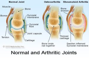
Active rheumatoid arthritis causes the joint to swell because of both thickening of the joint lining tissue (synovium) and because of excess joint fluid. The swollen, inflamed joint stretches and irritates the capsule that surrounds the joint. The joint capsule has nerves endings within it that immediately send pain signals to the brain.

Past rheumatoid arthritis can lead to permanent joint destruction with damaged cartilage, bone, and ligaments. When the damaged joint is used, it can cause intense pain.

Joint tenderness

Rheumatoid arthritis characteristically leads to tenderness of involved joints. This is because the inflamed joint lining tissue has irritated the nerves in the joint capsule. When the irritated joint capsule is compressed by external pressure, such as from touching the joint, it is frequently tender. The pain elicited from compression is immediate. This is one of the reasons that rheumatoid arthritis can lead to difficulty sleeping and insomnia.

Joint swelling

Swollen joints are very common in rheumatoid arthritis. Sometimes the joint swelling is minimal and can be difficult to appreciate. Other times the joint swelling is very apparent. Generally, people who are affected by rheumatoid arthritis can easily tell when their joints are swollen. The joint swelling can lead to loss of range of motion of the joint. Joint swelling in the fingers can make it hard to get rings off and on easily.

Joint redness

Redness occurs over joints when they are inflamed. The redness in the skin over an inflamed joint from rheumatoid arthritis occurs because the capillaries of that skin are widened by the adjacent inflammation. These widened capillaries are referred to as dilated capillaries.Joint redness does not occur in all inflamed joints from rheumatoid arthritis. Sometimes the inflammation in the joint is inadequate to cause the capillaries in the skin to dilate.

Joint warmth

Warmth of the joints affected by rheumatoid arthritis is a sign of active inflammation. Doctors look for joint warmth as they monitor the activity of the disease. As rheumatoid arthritis responds to treatment, joint warmth resolves. Sometimes joint warmth is present without visible joint swelling or redness.

Joint stiffness

Stiffness is a typical rheumatoid arthritis symptom. Joints that are affected by active rheumatoid arthritis are inflamed and characteristically stiffer in the morning than later in the day. Doctors use the duration of the morning stiffness as a measure of the severity of the active joint inflammation. As rheumatoid arthritis responds to treatment, the duration of the morning joint stiffness diminishes.

Loss of joint range of motion

As the joints of rheumatoid arthritis become more inflamed with active disease, they tend to have incomplete range of motion. The range of motion is limited by the swelling within the joint. This is typically associated with weakness in the involved areas.

Joints affected by longstanding rheumatoid arthritis commonly lose range of motion permanently.

Limping

Limping from poor lower extremity function can be caused by many diseases of the nerves, muscles, and bones of the lower extremities. Limping frequently occurs when rheumatoid arthritis affects the hips, knees, ankles, or feet. Pain, loss of range of motion, and joint swelling all can cause a person with rheumatoid arthritis to have a noticeable limp. It is not unusual for a young child with rheumatoid arthritis to have a painless limp as the first sign of the rheumatoid disease.

Joint deformity

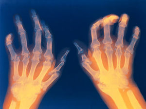
Joint deformity can occur from chronic rheumatoid arthritis. Deformity in rheumatoid arthritis occurs because the unchecked inflammation leads to both erosion of cartilage and bone as well as ligament loosening (laxity). Early detection and treatment of rheumatoid arthritis is critical to prevent permanent joint destruction and joint deformity.

Many joints affected (polyarthritis)

Usually, but not always, rheumatoid arthritis affects many joints. Classically, RA affects the small joints of the hands and wrists and balls of the feet. Also, not uncommonly, knees, elbows, hips, ankles, and shoulders can be inflamed.

Sometimes, only a few joints are involved. Less frequently, a singular joint is involved. Both of these scenarios are more common in childhood inflammatory arthritis (juvenile rheumatoid arthritis).

When four or more joints are inflamed, the condition is referred to as polyarthritis. When only a few joints are inflamed, it is referred to as oligoarthritis. When a single joint is inflamed, it is referred to as monoarthritis.

Both sides of the body affected (symmetric)

Typically, the distribution of the joints involved in a person with rheumatoid arthritis is similar on both sides of the body. This symmetric joint involvement is a feature of classic rheumatoid arthritis. This does not mean that joint involvement is always symmetric, but it is common.

Rheumatoid arthritis usually (not always) involves many joints on both sides of the body. It is, therefore, sometimes referred to as a symmetric polyarticular form of arthritis. Accordingly, the small joints of the hands, wrists, and feet are commonly affected. The knees, ankles, shoulders, hips, and elbows can also be involved in early disease. Rheumatoid arthritis is characterized by inflammation in these joints. Early manifestations of this inflammation can be gradual or rapidly intense. The joint inflammation causes stiffness, usually worse in the morning or after being sedentary. It also causes warmth, swelling, redness, and pain in varying degrees. The joint can be very subtly affected with slight swelling or markedly affected with substantial loss of range of motion. The pain level can be completely disabling and does not always correlate with the degree of apparent inflammation.

As described above, the manner that each of the symptoms affects an individual can be very different from individual to individual and can vary during the day. The intensity and effect of each of the symptoms is dependent upon the patient’s age, activity, the medications he or she takes, as well as any additional medical conditions that are present.

Loss of joint function

Because rheumatoid arthritis leads to pain, swelling, and tenderness of the involved joints, there is loss of joint function. The swelling and sensitivity impedes the full motion and stability of the joint and it becomes incapable of carrying the movement with confidence, balance, and completeness. This loss of joint function leads to limping, lack of coordination, loss of grip and dexterity, and disability.

Anemia

The chronic inflammation of rheumatoid arthritis commonly causes the bone marrow to decrease the release of red blood cells into the circulation. This lowers the red blood count to cause anemia when rheumatoid arthritis is active. It is not unusual for the anemia of rheumatoid arthritis to spontaneously correct as the inflammation of the disease is quieted by treatment.

Fever

Fever, while not common in rheumatoid arthritis, does occur in some patients when the disease is actively causing inflammation. Typically, there is only mild low-grade temperature elevation and this corrects rapidly as the inflammation of rheumatoid arthritis is treated. Because patients with rheumatoid arthritis frequently require medications that can decrease the normal immune response, it is important that when they develop fever, infection is considered as a possible cause. Infections can require aggressive treatment and interruption of some underlying rheumatoid treatments.

Diagnosis and stages of RA

There is no singular test for diagnosing rheumatoid arthritis. The diagnosis is based on the clinical presentation. Ultimately, rheumatoid arthritis is diagnosed based on a combination of the presentation of the joints involved, characteristic joint swelling and stiffness in the morning, the presence of blood rheumatoid factor (RF test or RA test) and citrulline antibody, as well as findings of rheumatoid nodules and radiographic changes (X-ray testing). It is important to understand that there are many forms of joint disease that can mimic rheumatoid arthritis.

The first step in the diagnosis of rheumatoid arthritis is a meeting between the health care professional and the patient. The doctor reviews the history of symptoms, examines the joints for inflammation, tenderness, swelling, and deformity, the skin for rheumatoid nodules (firm lumps or bumps under the skin, most commonly over the elbows or fingers), and other parts of the body for inflammation. Certain blood and X-ray tests are often obtained. The diagnosis will be based on the pattern of symptoms, the distribution of the inflamed joints, and the blood and X-ray findings. Several visits may be necessary before the health care professional can be certain of the diagnosis. A doctor with special training in arthritis and related diseases is called a rheumatologist.

It is the inflammation in the joint that helps to distinguish rheumatoid arthritis from common types of arthritis that are not inflammatory, such as osteoarthritis or degenerative arthritis. The distribution of joint inflammation is also important to the health care professional in making a diagnosis. In rheumatoid arthritis, the small joints of the hands and fingers, wrists, feet, and knees are typically inflamed in a symmetrical distribution (affecting both sides of the body). When only one or two joints are inflamed, the diagnosis of rheumatoid arthritis becomes more difficult. The doctor may then perform other tests to exclude arthritis due to infection or gout. The detection of rheumatoid nodules (described above), most often around the elbows and fingers, can suggest the diagnosis.

Abnormal antibodies can be found in the blood of people with rheumatoid arthritis with simple blood testing. An antibody called “rheumatoid factor” (RF) can be found in 80% of patients with rheumatoid arthritis. Patients with rheumatoid arthritis and rheumatoid factor are referred to as having “seropositive rheumatoid arthritis.” Patients who are felt to have rheumatoid arthritis and do not have positive rheumatoid factor testing are referred to as having “seronegative rheumatoid arthritis.” Citrulline antibody (also referred to as anti-citrulline antibody, anticyclic citrullinated peptide antibody, and anti-CCP antibody) is present in 50%-75% people with rheumatoid arthritis. It is useful in the diagnosis of rheumatoid arthritis when evaluating cases of unexplained joint inflammation. A test for citrulline antibodies is especially helpful in looking for the cause of previously undiagnosed inflammatory arthritis when the traditional blood test for rheumatoid arthritis, rheumatoid factor, is not present. Citrulline antibodies have been felt to represent the earlier stages of rheumatoid arthritis in this setting. Citrulline antibodies also have been associated with more aggressive forms of rheumatoid arthritis. Another antibody called the “antinuclear antibody” (ANA) is also frequently found in people with rheumatoid arthritis.

It should be noted that many forms of arthritis in childhood (juvenile inflammatory arthritis) are not associated with blood test positivity for rheumatoid factors. In this setting, juvenile rheumatoid arthritis must be distinguished from other types of joint inflammation, including plant thorn arthritis, joint injury, arthritis of inflammatory bowel disease, and rarely joint tumors.

A blood test called the sedimentation rate (sed rate) is a crude measure of the inflammation of the joints. The sed rate actually measures how fast red blood cells fall to the bottom of a test tube. The sed rate is usually faster (high) during disease flares and slower (low) during remissions. Another blood test that is used to measure the degree of inflammation present in the body is the C-reactive protein. Blood testing may also reveal anemia, since anemia is common in rheumatoid arthritis, particularly because of the chronic inflammation.

The rheumatoid factor, ANA, sed rate, and C-reactive protein tests can also be abnormal in other systemic autoimmune and inflammatory medical conditions. Therefore, abnormalities in these blood tests alone are not sufficient for a firm diagnosis of rheumatoid arthritis.

Joint X-rays may be normal or only demonstrate swelling of soft tissues early in the disease. As the disease progresses, X-rays can reveal bony erosions typical of rheumatoid arthritis in the joints. Joint X-rays can also be helpful in monitoring the progression of disease and joint damage over time. Bone scanning, a procedure using a small amount of a radioactive substance, can also be used to demonstrate the inflamed joints. MRI scanning can also be used to demonstrate joint damage.

The doctor may elect to perform an office procedure called arthrocentesis. In this procedure, a sterile needle and syringe are used to drain joint fluid out of the joint for study in the laboratory. Analysis of the joint fluid in the laboratory can help to exclude other causes of arthritis, such as infection and gout. Arthrocentesis can also be helpful in relieving joint swelling and pain. Occasionally, cortisone medications are injected into the joint during the arthrocentesis in order to rapidly relieve joint inflammation and further reduce symptoms.

What are the stages of rheumatoid arthritis?

The American College of Rheumatology has developed a system for classifying rheumatoid arthritis that is primarily based upon the X-ray appearance of the joints. This system helps medical professionals classify the severity of your rheumatoid arthritis with respect to cartilage, ligaments, and bone.

Stage I

No damage seen on X-rays, although there may be signs of bone thinning

Stage II

On X-ray, evidence of bone thinning around a joint with or without slight bone damage

Slight cartilage damage possible

Joint mobility may be limited; no joint deformities observed

Atrophy of adjacent muscle

Abnormalities of soft tissue around joint possible

Stage III

On X-ray, evidence of cartilage and bone damage and bone thinning around the joint

Joint deformity without permanent stiffening or fixation of the joint

Extensive muscle atrophy

Abnormalities of soft tissue around joint possible

Stage IV

On X-ray, evidence of cartilage and bone damage and osteoporosis around joint

Joint deformity with permanent fixation of the joint (referred to as ankylosis)

Extensive muscle atrophy

Abnormalities of soft tissue around joint possible

Rheumatologists also classify the functional status of people with rheumatoid arthritis as follows:

Class I: completely able to perform usual activities of daily living

Class II: able to perform usual self-care and work activities but limited in activities outside of work (such as playing sports, household chores)

Class III: able to perform usual self-care activities but limited in work and other activities

Class IV: limited in ability to perform usual self-care, work, and other activities

Treatment of RA

1. Conventional Medicine:

There is no known cure for rheumatoid arthritis. To date, the goal of treatment in rheumatoid arthritis is to reduce joint inflammation and pain, maximize joint function, and prevent joint destruction and deformity. Early medical intervention has been shown to be important in improving outcomes. Aggressive management can improve function, stop damage to joints as monitored on X-rays, and prevent work disability. Optimal RA treatment involves a combination of medications, rest, joint-strengthening exercises, joint protection, and patient (and family) education. Treatment is customized according to many factors such as disease activity, types of joints involved, general health, age, and patient occupation. RA treatment is most successful when there is close cooperation between the health care professional, patient, and family members.

Two classes of medications are used in treating rheumatoid arthritis: fast-acting “first-line drugs” and slow-acting “second-line drugs” (also referred to as disease-modifying antirheumatic drugs or DMARDs). The first-line drugs, such as aspirin and cortisone (corticosteroids [Rayos, Celestone, Depo-Medrol, Kenalog]), are used to reduce pain and inflammation. The slow-acting second-line drugs, such as methotrexate (Rheumatrex, Trexall, Otrexup, Rasuvo) and hydroxychloroquine (Plaquenil), promote disease remission and prevent progressive joint destruction.

The degree of destructiveness of rheumatoid arthritis varies among affected individuals. Those with uncommon, less destructive forms of the disease or disease that has quieted after many years of activity (“burned out” rheumatoid arthritis) can be managed with rest plus pain control and anti-inflammatory medications alone. In general, however, function is improved and disability and joint destruction are minimized when the condition is treated earlier with second-line drugs (disease-modifying antirheumatic drugs), even within months of the diagnosis. Most people require more aggressive second-line drugs, such as methotrexate, in addition to anti-inflammatory agents. Sometimes these second-line drugs are used in combination.

The areas of the body other than the joints that are affected by rheumatoid inflammation are treated individually. Sjögren’s syndrome can be helped by artificial tears and humidifying rooms in the home or office. Medicated eyedrops, cyclosporine ophthalmic drops (Restasis), are also available to help the dry eyes in those affected. Regular eye checkups and early antibiotic treatment for infection of the eyes are important. Inflammation of the tendons (tendinitis), bursae (bursitis), and rheumatoid nodules can be injected with cortisone. Inflammation of the lining of the heart and/or lungs may require high doses of oral cortisone.

In some cases with severe joint deformity, surgery may be recommended to restore joint mobility or repair damaged joints. Doctors who specialize in joint surgery are orthopedic surgeons. The types of joint surgery range from arthroscopy to partial and complete replacement of the joint. Arthroscopy is a surgical technique whereby a doctor inserts a tube-like instrument into the joint to see and repair abnormal tissues.

Total joint replacement is a surgical procedure whereby a destroyed joint is replaced with artificial materials. For example, the small joints of the hand can be replaced with plastic material. Large joints, such as the hips or knees, are replaced with metals.

2. Traditional Chinese Medicine

Quickly Relieve Your Pains, Stiffness, and Sufferings from Ankylosing Spondylities (AS) or Rheumatoid Arthritis (RA). Firmly STOP the progression of Your Conditions (Ankylosing Spondylities or Rheumatoid Arthritis). So far over 250,000 RA and AS patients have regained their health and they can live and work normally with no pains, no swells, and no more medicines.

Our Natural Treatment is both Effective(see the witnesses below) and Simple to Apply. (1)You clearly tell us the

detailed History of both Diagnosis and Treatments of Your conditions (All medicial records). If neccessary, we will do video diagnosis with you. (2)Our Doctor team will formulate the herb recipe specially for You – All of the herbs are quality-sured. We will express them to You (3)You take it AT YOUR HOME under the instructions of our Doctors.

What is Ankylosing Spondylitis?

Ankylosing spondylitis or AS, is a form of arthritis that primarily affects the spine, although other joints can become involved. It causes inflammation of the spinal joints (vertebrae) that can lead to severe, chronic pain and discomfort.

In more advanced cases this inflammation can lead to ankylosis — new bone formation in the spine — causing sections of the spine to fuse in a fixed, immobile position.This fusing makes the spine less flexible and can result in a hunched-forward posture.

AS can also cause inflammation, pain, and stiffness in other areas of the body such as the shoulders, hips, ribs, heels, and small joints of the hands and feet. Sometimes the eyes can become involved (known as iritis or uveitis), and — rarely — the lungs and heart can be affected. If ribs are affected, it can be difficult to breathe deeply.

Ankylosing spondylitis affects men more often than women. Signs and symptoms typically begin in early adulthood.

The hallmark feature of ankylosing spondylitis is the involvement of the sacroiliac (SI) joints during the progression of the disease. The SI joints are located at the base of the spine, where the spine joins the pelvis.

There is no cure for ankylosing spondylitis, but treatments can lessen your symptoms and possibly slow progression of the disease.

Causes of Ankylosing Spondylitis

Ankylosing spondylitis has no known specific cause, though genetic factors seem to be involved. In particular, people who have a gene called HLA-B27 are at greatly increased risk of developing ankylosing spondylitis. However, only some people with the gene develop the condition. Most individuals who have AS also have a gene that produces a “genetic marker,” a protein called HLA-B27. This marker is found in more than 95 percent of people in the Caucasian population with AS. It is important to note, however, that one does not have to be HLA-B27 positive to have AS. Also, a majority of people with this marker never develop ankylosing spondylitis.

Scientists suspect that other genes — along with a triggering environmental factor such as a bacterial infection, for example — are needed to activate AS in susceptible people. HLA-B27 likely accounts for about 30 percent of the overall risk, but there are numerous other genes working in concert with HLA-B27. Researchers have identified more than 60 genes that are associated with AS and related diseases. Among the newer key genes identified are ERAP 1, IL-12, IL-17, and IL-23.

One classic hypothesis has been that AS may start when the defenses of the intestines break down and certain bacteria pass into the bloodstream, triggering changes in the immune response. The association between ankylosing spondylitis and HLA-B27 varies greatly between ethnic and racial groups.

Risk factors:

The risk factors that predispose a person to AS include:

Testing positive for the HLA-B27 marker

A family history of AS

Frequent gastrointestinal infections

Unlike other forms of arthritis and rheumatic diseases, general onset of AS commonly occurs in younger people, between the ages of 17 and 45. However, it can also affect children and those who are much older.

Symptoms and Complications of AS

The severity of AS varies greatly from person to person, and not everyone will experience the most serious complications or have spinal fusion. Some may experience only intermittent back pain and discomfort, while others may experience severe pain and stiffness over multiple areas of the body for long periods of time. AS can be debilitating and, in some cases, lead to disability.

Early signs and symptoms of ankylosing spondylitis might include pain and stiffness in your lower back and hips, especially in the morning and after periods of inactivity. Neck pain and fatigue also are common. Over time, symptoms might worsen, improve or stop at irregular intervals.

The areas most commonly affected are:

The joint between the base of your spine and your pelvis (sacroiliac)

The vertebrae in your lower back

The places where your tendons and ligaments attach to bones (entheses),mainly in your spine, but sometimes along the back of your heel

The cartilage between your breastbone and ribs

Your hip and shoulder joints

Almost all cases of AS are characterized by acute, painful episodes (also known as “flares”), which are followed by temporary periods of remission when symptoms subside.

It is important to know that ankylosing spondylitis is a chronic, or lifelong, disease and that the severity of AS has nothing to do with age or gender. It can be just as severe in women and children as in men.

Remember that even if you have AS and are experiencing only mild symptoms, which you are able to manage well, it is important to see your rheumatologist once a year in order to detect and treat any underlying complications.

Complications:

In severe ankylosing spondylitis, new bone forms as part of the body’s attempt to heal. This new bone gradually bridges the gap between vertebrae and eventually fuses sections of vertebrae. Those parts of your spine become stiff and inflexible. Fusion can also stiffen your rib cage, restricting your lung capacity and function.

Other complications might include

Eye inflammation (uveitis). One of the most common complications of ankylosing spondylitis, uveitis can cause rapid-onset eye pain, sensitivity to light and blurred vision. See your doctor right away if you develop these symptoms.

Compression fractures. Some people’s bones thin during the early stages of ankylosing spondylitis. Weakened vertebrae can crumble, increasing the severity of your stooped posture. Vertebral fractures can put pressure on and possibly injure the spinal cord and the nerves that pass through the spine.

Heart problems. Ankylosing spondylitis can cause problems with your aorta, the largest artery in your body. The inflamed aorta can enlarge to the point that it distorts the shape of the aortic valve in the heart, which impairs its function.

Diagnosis of Ankylosing Spondylitis

The diagnosis of ankylosing spondylitis is based on several factors, including:

Findings of a physical exam

In the physical exam, to bend in different directions to test the range of motion in your spine. To press on specific portions of your pelvis or by moving your legs into a particular position to reproduce your pain. To take a deep breath to see if you have difficulty expanding your chest,etc…

Imaging tests

X-rays allow your doctor to check for changes in your joints and bones, though the visible signs of ankylosing spondylitis might not be evident early in the disease.

An MRI uses radio waves and a strong magnetic field to provide more-detailed images of bones and soft tissues. MRI scans can reveal evidence of ankylosing spondylitis earlier in the disease process, but are much more expensive.

Lab tests

There are no specific lab tests to identify ankylosing spondylitis. Certain blood tests can check for markers of inflammation, but inflammation can be caused by many different health problems.

Your blood can be tested for the HLA-B27 gene, but most people who have that gene don’t have ankylosing spondylitis, and fewer black people with the disease have the gene than do white people.